
 Oral & Maxillofacial Surgery
Beating Cheek Cancer Without Losing His Face: A Microvascular Reconstruction Story
Oral squamous cell carcinoma involving the inner lining of the right cheek. His greatest fear was not the cancer itself, but waking up unable to open his mouth properly, with a sunken cheek and visible scarring across his face.
Patient
Mr. Sunil, age 61
Timeline
7-hour single-stage surgery; 8–10 day hospital stay; 4–6 weeks for major healing; ongoing rehabilitation through months 2–3
Treatment
Oral & Maxillofacial Surgery
Treatment Approach
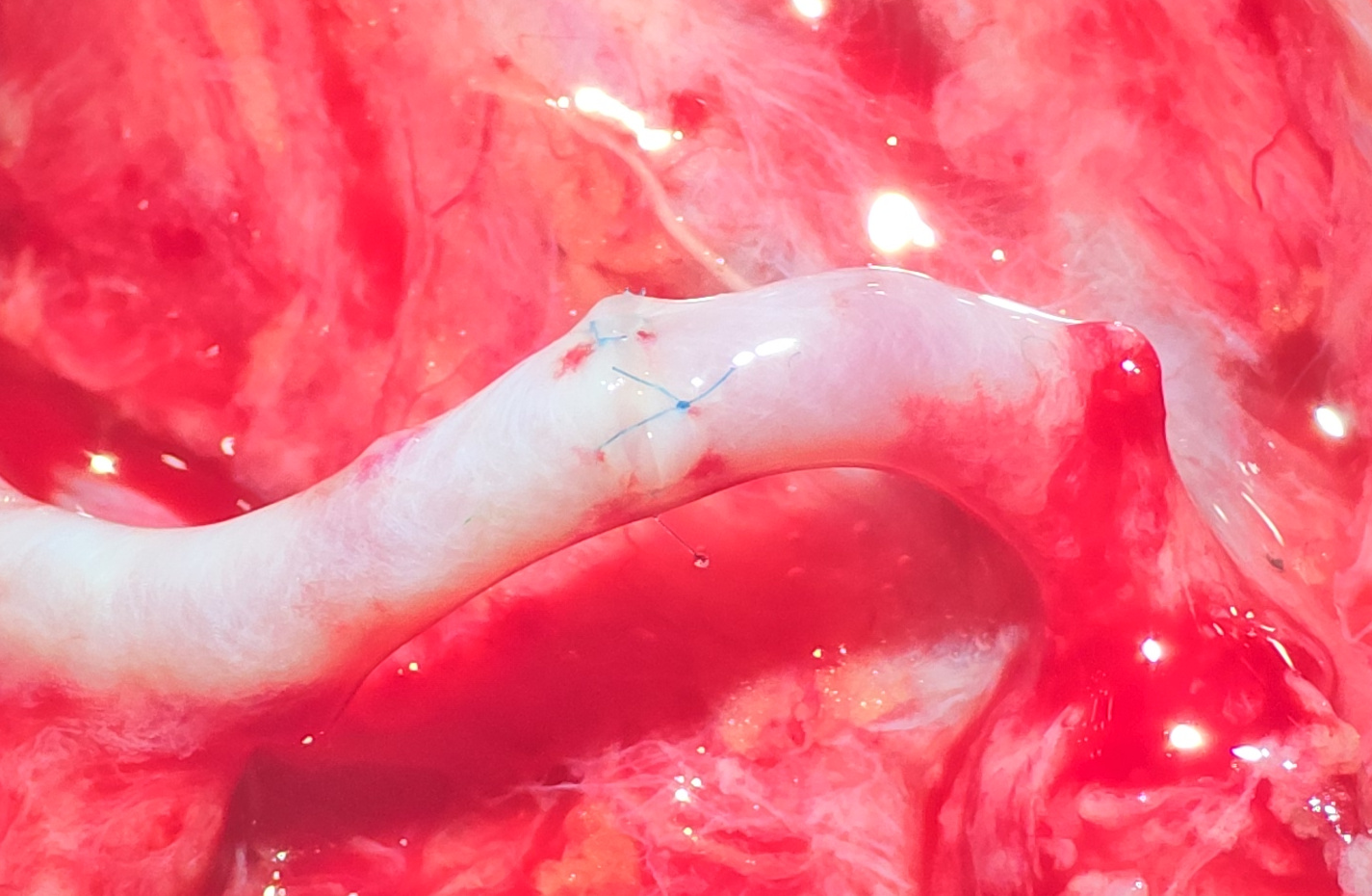
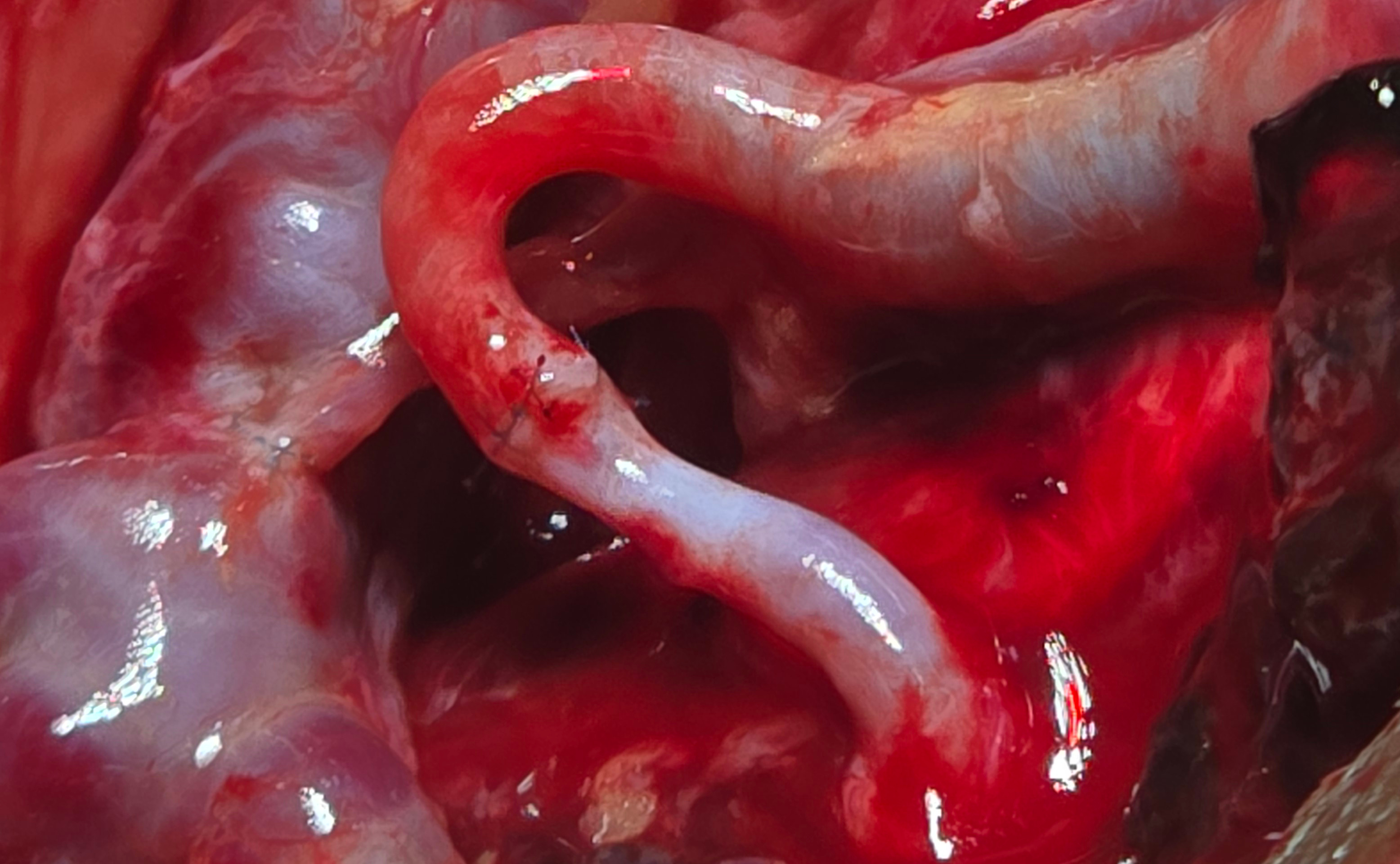
A single-stage 7-hour procedure led by Dr. Manjula Herath: wide local excision of the buccal tumour with clear margins and selective neck dissection, followed by reconstruction using a radial forearm fasciocutaneous free flap. Tissue (skin, fascia and the radial artery with its accompanying veins) was harvested from the left forearm and transferred to rebuild the inner cheek. Microsurgical anastomoses (1 artery + 2 veins, 2–3 mm in diameter) were performed under an operating microscope using sutures finer than human hair, connecting the flap's vessels to the facial artery and a neck vein. Continuous Doppler monitoring was used postoperatively to confirm blood flow to the flap.
Mr. Sunil’s Story
Microvascular reconstruction is one of the most advanced procedures in oral and maxillofacial surgery. It involves transferring living tissue from one part of the body to rebuild a missing area, with blood vessels as small as 1–2 mm reconnected under an operating microscope so that the transplanted tissue stays alive from the first moment. At 61, Mr. Sunil was diagnosed with oral squamous cell carcinoma involving the inner lining of his right cheek. He had noticed a non-healing ulcer for months before seeking advice, and by the time he was referred his greatest fear was not the cancer itself but the surgery — he had heard that removing a tumour of this size would leave him unable to open his mouth properly, with a sunken cheek and visible scarring across his face. Dr. Manjula Herath proposed a single-stage procedure: remove the cancer with clear margins through the mouth where possible, and immediately rebuild the defect using a radial forearm free flap — the gold standard for soft-tissue oral cavity reconstruction because of its thin, pliable, highly vascular tissue. The surgery took seven hours. Two surgical teams worked in parallel — one removing the tumour with safe margins and clearing the neck nodes, the other harvesting the forearm flap with its vascular pedicle. Once the tumour was out, the flap was brought up to the face, inset to recreate the inner cheek lining, and its vessels connected to the neck under the microscope. The first Doppler signal after release of the clamps confirmed perfect blood flow. The reconstruction was a complete success. Speech and mouth-opening exercises began within the first week; soft foods returned within weeks; and by the end of the second month Mr. Sunil was eating, speaking, and chewing on the reconstructed side normally. "I was so afraid I would not look like myself," he said. "My family says my face looks the same. I can speak, I can eat, and the cancer is gone. Dr. Herath did not just save my life — he gave me back the face I came in with."
The Outcome
Cancer removed with clear margins. The flap was 100% successful. Mouth opening, speech, and swallowing function preserved. Mr. Sunil is back to enjoying meals with his family, with intact facial contour and minimal change to his appearance.
“I was so afraid I would not look like myself. My family says my face looks the same. I can speak, I can eat, and the cancer is gone. Dr. Herath did not just save my life — he gave me back the face I came in with.”
— Mr. Sunil
Treating Consultant
Dr. Manjula Herath
Oral & Maxillofacial Surgery
Related Services
Ready to start your journey?
Book a consultation to discuss your treatment options with our specialists.